Prediction of preeclampsia
Measurement of uterine artery PI (UTPI)
The UTPI can be measured by either transabdominal or transvaginal sonography.
For the transabdominal scan in the first trimester, a sagittal section of the uterus should be obtained and the cervical canal and internal cervical os identified. The transducer should be gently tilted from side to side and color Doppler should be used to identify each uterine artery along the side of the cervix and uterus at the level of the internal os.
- In the second and third trimesters, color Doppler should be used to identify each uterine artery at the apparent crossover with the external iliac arteries.
For the transvaginal scan, women should be asked to empty their bladders and placed in the dorsal lithotomy position. The ultrasound probe should then be inserted into the vagina and placed in turn into the left and right lateral fornix. The uterine arteries are identified using color Doppler at the level of the internal cervical os.
After identification of each uterine artery, pulsed wave Doppler should be used with the sampling gate set at 2 mm to cover the whole vessel. Care should be taken to ensure that the angle of insonation is less than 30º. It is important that the peak systolic velocity is greater than 60 cm/s to ensure that the uterine artery, rather than the arcuate artery, is being examined.
When three similar consecutive waveforms are obtained the PI should be measured and the mean PI of the left and right arteries calculated.
Pulsatility index = (Peak systolic velocity - minimum diastolic velocity) / Mean velocity
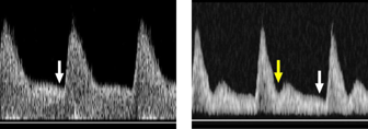
The left waveform has good end-diastolic flow (white arrow). The right waveform shows high resistance of flow with early diastolic notch (yellow arrow) and low end-diastolic flow (white arrow).